Below is a summary of the abstract you submitted. Presenting author(s) is shown in bold.
If any changes need to be made, you can modify the abstract or change the authors.
You can also download a .docx version of this abstract.
If there are any problems, please email Dan at dar78@pitt.edu and he'll take care of them!
This abstract was last modified on March 8, 2022 at 4:59 p.m..


Bacteriophages, bacteria-infecting viruses, are very abundant across the world and have the potential to create numerous advancements in the fields of biology and medicine. From creating an alternative to antibiotics as well as other therapeutics such as CRISPR gene modification, phage research is an important field of study. Keeping this in mind, we followed the HHMI SEA-PHAGES protocols in the hopes of finding phages, isolating their DNA, analyzing it, and overall contributing to the database of known phages.
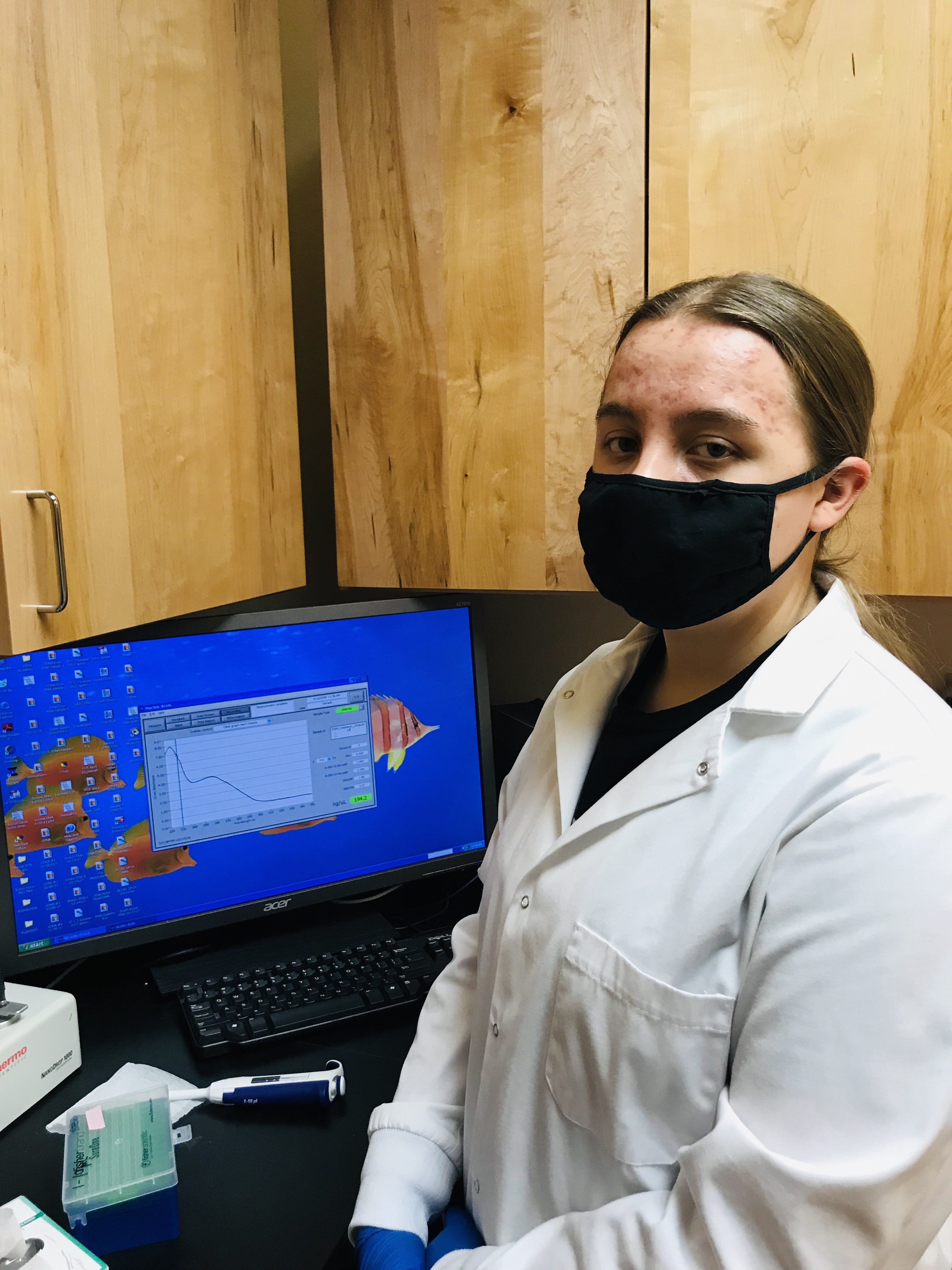
Using microbiology techniques, we isolated a single phage type, amplified it to a usable concentration, and properly prepared it for genome sequencing. The first set of procedures we followed had to do with isolation, using enriched isolation with the host bacterium M. Smegmatis rather than direct. Next, we purified our phage in order to have one phage type. We then amplified our phage to obtain a high-titer lysate and were then ready for the final steps: imaging our phage using transmission electron microscopy; archiving the phage for later use; extracting the DNA and using the NanoDrop spectrophotometer to find its quality and quantity; and finally characterizing the phage genome using restriction enzymes and gel electrophoresis.
Out of six original soil samples, we successfully derived phage from one sample located under a water faucet in Polson, MT. After we isolated and amplified phages from this sample, we calculated the titer of our concentrated solutions. Our high-titer samples were then sent to the University of Pittsburgh for genome sequencing. The results indicated that the two samples sequenced were identical. In addition to the genome sequencing, we performed a restriction enzyme digest and electrophoresis, creating DNA fingerprints for our phage samples. The results were inconclusive, however. Only one of the three fingerprints showed any bands in the gel. The ladder worked as intended, the uncut DNA had a single distinct band, the DNA treated with the enzyme HindIII showed a smear throughout the column, but the other enzymes showed no results.
We successfully isolated, purified, amplified, and characterized our phage. We hoped to get three unique phages, but only ended up with one, GoldenAsh and Voidmere being identical. We viewed the GoldenAsh genome map on Phamerator, and plan on annotating the phage genome to determine the gene locations and functions. Once fully annotated, it can be compared to other phage genomes. During this course, our team has learned how to use a variety of lab techniques related to microbiology. In hindsight, we could have done some things better. If we were to conduct another experiment or study we would extract samples from geographically different locations, as similar locations may yield similar phage.